The winning pictures in the DNRF’s photo competition 2019 have been chosen
Photos have the ability to unseal the world of science in a surprising and inviting way by revealing its beauty or otherwise fascinating appeal. The DNRF would like to share with a broader audience how scientific discovery each day advances our knowledge of ourselves and the world we live in. To this end, and for the second time, the foundation has launched a photo competition based on the photograph’s potential as documentation and communication of scientific research.
Below, you can see the winning pictures and find links to interviews with the winners, who explain the fascinating research behind the pictures.
-
Selection criteria: - Degree to which the photo evokes emotions in the observer
- Degree to which the photo works as visual entry point to the story behind the specific research result
- Aesthetic quality of the photo
Panel: - Christine Buhl Andersen, Director at Ny Carlsberg Glyptoteket
- Louise Wolthers, Research Manager/Curator at the Hasselblad Foundation
- Minik Rosing, Professor at the Natural History Museum, board member at the DNRF and Louisiana Museum of Modern Art
First prize: Two root nodules. Niels Sandal, Senior researcher, Aarhus University

The panel’s review: Two root nodules by Niels Sandal. The photo is magical and not immediately open to interpretation. It is not possible to see whether the scale is at a cellular level or a cluster of galaxies, but the observer is drawn to the world that opens up in the picture. Further, the photo is very beautiful and well composed, and it gives a sense of movement and space. The scientific content is important as it shows how the scientist works with the symbiosis between bacteria and plants, which thereby can grow without addition of nitrogen fertilizer.
Read the interview with Niels Sandal about the science behind the photo here
Second prize: A hedgehog getting a dental examination. Sofie Lund Rasmussen, Ph.D student, University of Southern Denmark and Naturama
The panel’s review: “A hedgehog getting a dental examination” by Sofie Lund Rasmussen. The photo exudes horror in contrast to the cute hedgehog we all know from the garden. There is no technological filter between the motif and the observer. It infuses respect for nature and its creatures. The photo illustrates research on the familiar nature driven by care for wild animals, here the dental health of wild hedgehogs. It illustrates both scientific curiosity and empathy with the animals we live among.
Read the interview with Sofie Lund Rasmussen about the science behind the photo here
Third prize: Brain cancer cell on the go. Johann Mar Gudbergsson, Ph.D. student Aalborg University
-
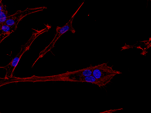
Brain cancer cell on the go, Johann Mar Gudbergsson, Aalborg University
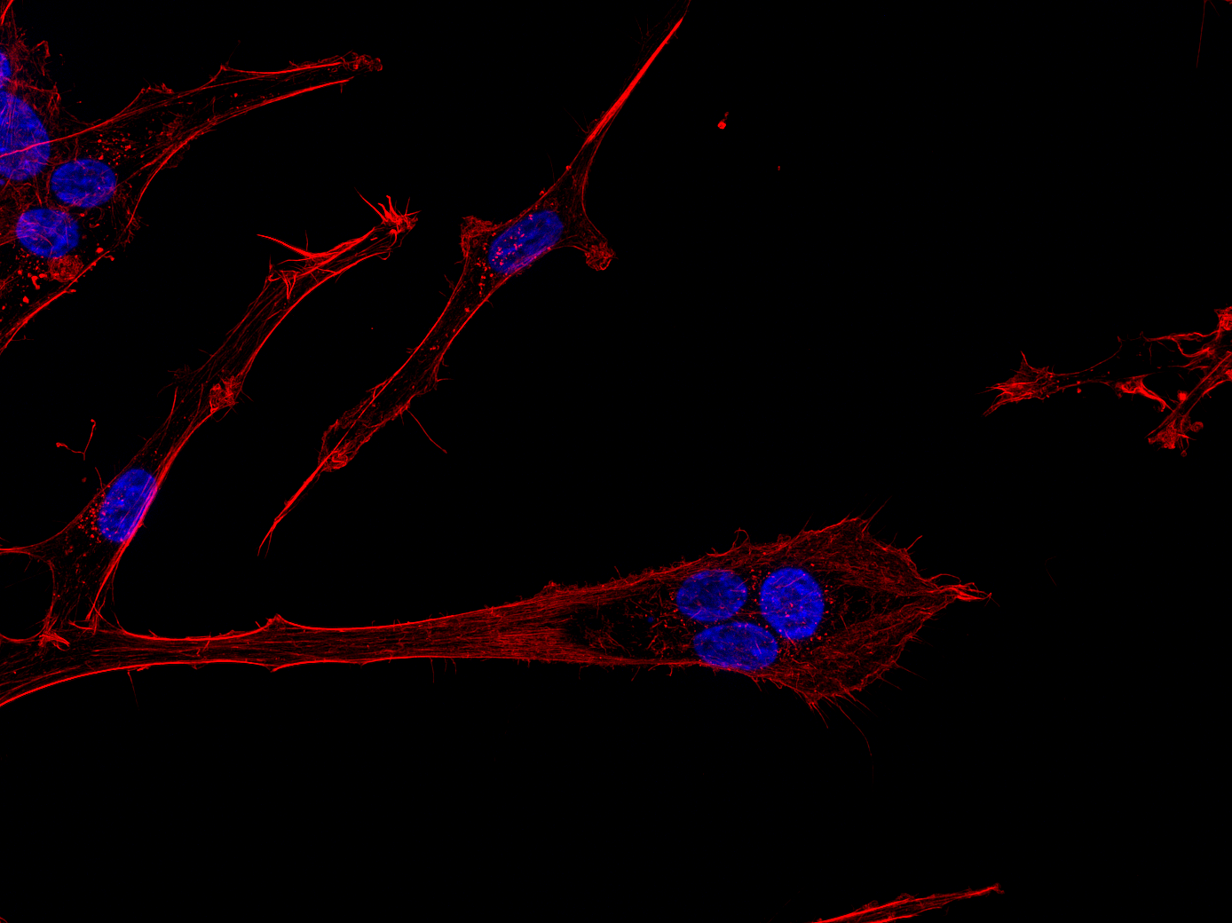
The panel’s review: “Brain cancer cells on the go” by Johann Mar Gudbergsson. The photo has a visual beauty that holds a sense of movement that underpins the invasive character of the depicted cancer cells. There is a demonic dimension in the motif that communicates the seriousness of the disease and, in that, the importance of the researcher’s work.
Read the interview with Johann Mar Gudbergsson about the science behind the photo here
Media partners:

![]()
See some of the pictures from this year's competition in the gallery below
. I use immunofluorescent microscopy to follow different components of cancer cells after treatment and study how they react. The more we can see – the more we know.")

 og forskellige bakterietyper (lilla og grønne celler), der koloniserer på en tand med caries. Forskningen giver ny viden om, hvordan svampe lever og interagerer med bakterier og hvordan dette samspil potentielt kan påvirke miljøet i tandbelægningen. Der er brugt en molekylærbiologisk teknik kaldet ”fluorescens in situ hybridisering”, der giver mulighed for at identificere de forskellige mikroorganismer i naturlige tandbelægninger ved hjælp af fluorescens mikroskopi.")

, and the plant’s cell nuclei are colored with DAPI. The nuclei are seen as light blue dots. You can see the bacteria in the young nodule and on the surface of the older nodule. In our research group, we have isolated many of the plant genes that are necessary for symbiosis.")
 can show different microstructure when it is treated differently. Thus, can be applied for different purposes. The left side (fall leaves structure) is produced when the graphite is simply polished with normal A4 paper. It has low capacitive current which is useful to immobilize DNA for genosensor. The right (rocky structure) is generated when the graphite is polished with sand paper, producing many edge planes which is best for protein immobilization for biosensor application. This image is taken when I did my PhD at Interdiscpinary Nanoscience Center (iNANO), Aarhus University, Aarhus-Denmark.")

 and actin cytoskeleton (red). The cell in the center of the photo is polynuclear, meaning that it contains multiple nuclei and therefore more DNA, which is often associated with cancer stem cells. Those are quite happy to move around, which can be seen in the photo by the long cellular process that stretches away from the blue nuclei. This shows the directions in which the cancer cell is heading. In patients with this type of brain cancer, it is the invasive cancer cells that constitutes a huge problem, because, with current treatments, they are impossible to exterminate. By invading healthy brain tissue, they are capable of using the brain’s own protection mechanisms, which, among other things, prevent harmful substances from entering the brain, and thereby also part of the compounds available for treatment.")
 imaging of brain cancer spheroid models. The sample is nucleic acid stained and not fixed - which allows for dynamic measurements – and we obtain single-cell resolution on the length scale of an entire tumor model (up to 1 mm).")