The winning pictures in the DNRF’s photo competition 2018 have been chosen
This year, the Danish National Research Foundation launched a photo competition for the foundation’s grantees, and now the winners have been chosen. Below, you can see the winning pictures and find links to interviews with the winners, who explain the fascinating research behind the pictures.
The selection criteria:
- Degree to which the photo raises curiosity in the eye of the beholder
- Degree to which the photo works as a visual entry point to the story behind the specific research result
- Aesthetic quality of the photo
The selection panel: Christine Buhl Andersen, Director at Glyptoteket; Louise Wolthers, Research Manager/Curator at the Hasselblad Foundation; and Minik Rosing, Professor at the Natural History Museum and board member at the DNRF and the Louisiana Museum of Modern Art
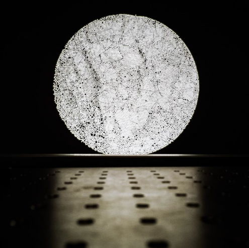
First Prize: The rising fiber moon, Senior Researcher Jonas Schou Neergaard-Nielsen from the Center for Macroscopic Quantum States (bigQ)

The panel’s review: The photo’s beauty and photographic quality make it immediately fascinating. What at first glance looks like the full moon in a night sky turns out to be a wall projection of microscopic fibers. The photo shows an innovative use of microscopy. In terms of quality, the photo is perfect with regard to composition, lighting, and distinctness. It is a classic photo and, at the same time, an advanced high-tech experimental display.
Read the interview with Jonas Schou Neergaard-Nielsen about the science behind the photo here
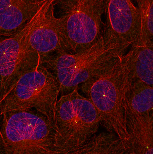
Second Prize: The nests, Post-doc Søs Grønbæk Mathiassen from the Center for Autophagy, Recycling and Disease (CARD)

The panel’s review: This photo is a beautiful composition in both structure and color, which speaks to imagination and curiosity. It makes the observer want to find out what the photo shows. It creates a number of associations with jellyfish, nebula, and organs, but the scale and material are unclear. The photo is fascinating in terms of understanding what autophagy is and how fluorescent photo micrography works.
Read the interview with Søs Grønbæk Mathiassen about the sicence behind the photo here
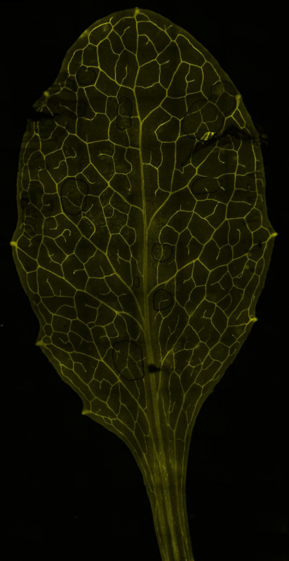
Third Prize: Confocal laser-scanning photomicrograph of a cress leaf, Ph.D. Fellow Pascal Hunziker from the Center for Dynamic Molecular Interactions (DynaMo)

The panel’s review: A beautiful and poetic picture of something as recognizable as a leaf appears in a whole new light in this photo. The delicate and fragile structures are actually part of the plant’s defense system, which has been made visible by advanced protein markers. The photo has a high technical quality and looks like a harmonious whole, even though it is composed of 74 smaller pictures covering 15 layers of the leaf’s depth.
Read the interview with Pascal Hunziker about the science behind the photo here
