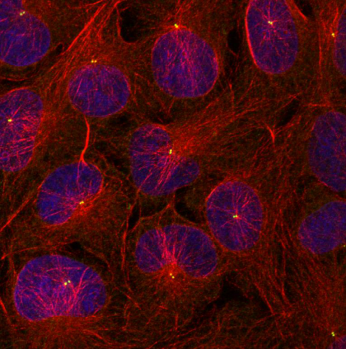
DNRF’s photo competition 2018: Close-up of cancer cells’ interphase wins second prize
A picture of cancer cells in the phase before cell division has won second prize in the Danish National Research Foundation’s photo competition 2018. The picture is part of a research project at the Center for Autophagy, Recycling and Disease (CARD), where post-doc Søs Grønbæk Mathiassen has examined how the cells’ embedded trashcan can influence the cancer cells’ ability to divide themselves.

At first glance, the design looks like an abstract work of art. Long threads in red shades run criss-cross in a complicated pattern, and the network connects a couple of almost circular forms in blue and purple, each of which has two illuminated dots placed close to each other. This beautiful picture has won second prize in the Danish National Research Foundation’s photo competition 2018, in which all of the foundation’s grant holders had the opportunity to submit fascinating pictures from the world of basic research that awaken our curiosity. The abstract pattern in this photo is actually a picture of cancer cells as seen through a microscope.
“The red threads are so-called microtubules, which are long thin pipes made up of the protein tubulin. They provide the cells with structure and function as a transportation system in the cells. The blue and purple areas are the nucleus – a membrane structure that holds the cells’ genetic material. And the small illuminated green dots are so-called centrosomes, which are small organelles in the cell, whose main purpose is to create these networks of microtubules. Each pair of dots, placed near each other, constitutes one centrosome,” explained the creator of the picture, Søs Grønbæk Mathiassen, a post-doc at the Center for Autophagy, Recycling and Disease (CARD) at the Danish Cancer Society.
Mathiassen has examined the cells’ ability to divide themselves
In the picture, the cancer cells are in the part of their life cycles called the interphase – a long preparatory phase during which the cells’ content and DNA duplicate in order to prepare for the phase called mitosis, which is the actual cell division. Mathiassen’s research has focused on centrosomes because they play an important role in cell division. They regulate how the genetic material – the DNA – is being divided. Therefore, it is important that the centrosomes function as intended so that the cell can divide itself correctly.
“During mitosis, two centrosomes almost create a web of microtubules between them and pull the DNA in two, to divide it, so to speak. In that way, the centrosomes and microtubules make sure that the DNA is divided correctly, but with cancer cells, you oftentimes see mistakes in the centrosomes, or that there are simply too many centrosomes,” Mathiassen explained.
In the winning photo, the centrosomes look like they are supposed to in that part of the cells’ life cycle, and the sample therefore constitutes an excellent foundation on which to compare other cell samples that are manipulated by the researchers. For instance, in the research project, Mathiassen adjusted the cells’ so-called autophagy both up and down in order to see how this mechanism affects and regulates the centrosomes.
Autophagy is the mechanism that the cell uses to decompose its own components to reuse them to get nutrition and energy – a kind of revamp that helps to repair and maintain the cell. The process therefore influences many cellular functions, and different extrinsic factors, including stress and lack of nutrition, can regulate the cells’ autophagy either up or down.
“With autophagy, damaged parts such as proteins and mitochondria, which can be toxic for the cell, are being decomposed and recycled. That is why autophagy is also called the “trashcan” of the cell, since it prohibits toxic waste from piling up,” Mathiassen said.
CARD studies the cells’ survival mechanism
Mathiassen’s project on centrosomes is one of CARD’s many research projects that, to some extent, study autophagy and how it affects cancer cells. Because just as autophagy helps healthy cells stay healthy and divide themselves correctly, the process also helps cancer cells survive when cancer is first established.
“In reality, autophagy is just a survival mechanism for the cell, no matter the cell type, and that is why autophagy will help the cancer cells to survive and become multifarious. Cancer cells can actually become addicted to the mechanism because the cell type has a high metabolism and therefore needs a lot of nutrition to keep dividing itself,” Mathiassen explained. He added:
“In that sense, autophagy is very important when it comes to cancer research and our knowledge about the exact role of the mechanism in the establishment and development of the cancer cells and how autophagy interferes with the cells’ other mechanisms and components, such as centrosomes, is still relatively small.”
The projects at CARD involve everything from a tiny Petri dish to small molecular organisms and bigger animal models in which autophagy is regulated. This all helps to create the best understanding possible of how the cells’ many complicated processes function and how they affect the body.
