The picture of a contaminated cross-section sample wins 3rd prize in the DNRF’s Photo Competition 2022
The winner of the third prize in the DNRF’s Photo Competition 2022 is the picture of a cross-section of a sample with different super-lattice structures along with some “dirt” on the surface. The image was discovered during a routine check at the Center of Excellence Center for Quantum Devices (QDev) at the University of Copenhagen.
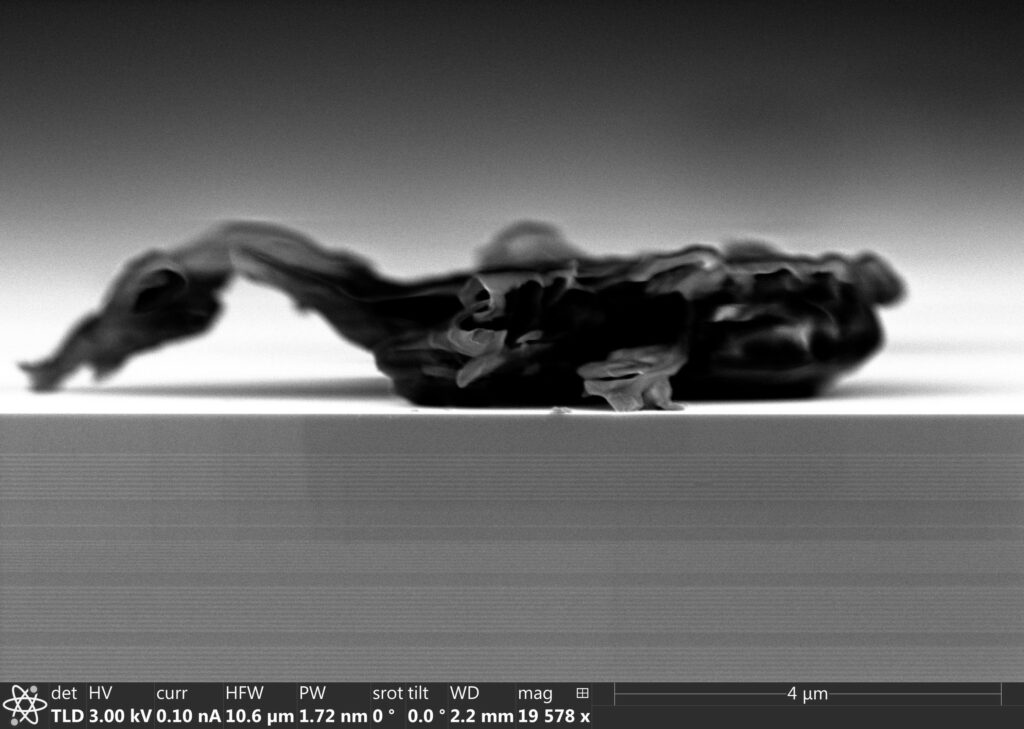
The picture captures a grey image that resembles an animal, yet not something that we have ever seen before. The blurry and complex expression of the image makes it hard to define. The winner of the 3rd prize in the DNRF’s Photo Competition 2022 is the picture of the cross-section of a sample and was taken by Ph.D. fellow Steffen Zelzer from the Center of Excellence Center for Quantum Devices (QDev), who discovered it during a routine check at the University of Copenhagen.
“It shows a scanning electron image of the cross-section of a sample with different super-lattice structures (the horizontal grey lines) and some “dirt” on the surface of the sample that looks very much like an animal from another world,” said Zelzer.
Potential to revolutionize communication and information processing
QDev studies how to create, measure, control, and protect quantum coherence and entanglement in solid-state electronic devices. The goal is to one day bring the entanglement under control and make it a resource, since it has the potential to revolutionize communication and information processing.
The image was taken during a routine check and shows a test sample that has been contaminated, which usually doesn’t happen.
“We discovered this during a routine check of how well we can cleave these samples that are grown in a molecular beam epitaxy chamber on indium arsenide wafers with layers of gallium antimony (the brighter lines),” said Zelzer.
The image shows a cross-section, which appears when the researchers break a sample in half and then are able to expose the cross-section.
Not an everyday occurrence
The research behind this picture started out as a collaboration between the University of Copenhagen and Microsoft, which has since ended. Zelzer wished to continue the work and to use it in his thesis as proof of concept, since part of his Ph.D. was about the design of a setup to cleave or break samples in an ultra-high vacuum.
“The image I sent in to the competition was additional information we gathered afterwards to show the ‘larger scale’ surface structure, because scanning tunneling microscopy is very shortsighted, meaning the maximal area one can observe is only a few micrometers at a time, but we were interested in what was the surface quality in the range of a few micrometers to half a millimeter – which is the thickness of the sample,” Zelzer said.
The surface is usually clean, so when the image appeared with some “dirt” on it wasn’t an everyday occurring.
The end of the sample’s life
The sample is part of a larger sample, but this sample’s life ended with this picture. The research behind the sample is very basic but is aimed at improving the final structure, which will then show the particles the researchers are looking for.
“I think the image shows what fascinates me about science: that you sometimes can start a seemingly mundane task of checking the quality of a sample and encounter the most beautiful and surprising results. It also shows humanity’s ability to see life in the form of shapes that remotely remind us of a face,” said Zelzer.
Zelzer is currently finishing his Ph.D. and working on his thesis.