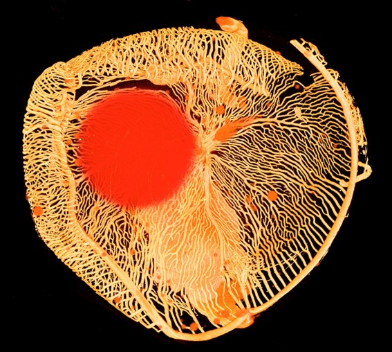
A scan of the eye of an Antarctic icefish wins third prize in the DNRF’s Photo Competition 2020
-
The three winning photographs in the Danish National Research Foundation’s Photo Competition 2020 have been found. In a series of three articles, the winners talk about their images and the research behind them. This article is about the third place winner, which this year went to Henrik Lauridsen, assistant professor in the Department of Clinical Medicine at Aarhus University and Jesper Skovhus Thomsen, associate professor in the Department of Biomedicine at Aarhus University. The article about the first place winner can be found here and the article about the second place winner here. More photos and information about the competition can be found here.
-
The panel’s review:
The scanned icefish eye creates a spatiality with a high degree of materiality and structure. The picture is at once decorative and full of scientific information. The shredded structures indicate the sensitivity
of the eye and the red circle adds mystery to the picture.The Panel:
- Christine Buhl Andersen, Chair of the New Carlsberg Foundation
- Louise Wolthers, Research Manager/Curator at the Hasselblad Foundation
- Minik Rosing, Professor at GLOBE Institute, Vice Chair of the DNRF board and board member at Louisiana Museum of Modern Art
The Antarctic icefish is a distinctive animal with milky white blood and the densest network of blood vessels in the eye’s retina known in any animal. A scanned photo of this network in the fish’s eye has won third prize in the foundation’s Photo Competition 2020. The research behind the photo dives into both the evolution of the eye through millions of years across vertebrates and into the peculiar biology of the ice fish.

An intricate network of elongated tracks that crisscross each other light up on a black background. To the left, at the top of the picture, one can see a round reddish spot that evokes memories of the sun on a summer night. This is the subject of the photo, that took third prize in the Danish National Research Foundation’s Photo Competition 2020, in which researchers from all over Denmark had the chance to send their best research photos. Even though the picture looks like abstract art, it is actually an example of nature’s wondrous design.
“The picture actually shows a scan of the eye of an Antarctic icefish. This network of branches is small blood vessels, which are located on the inside of the retina in the eye of the fish, and the red spot, slightly oblique to the left, is the lens of the eye,” said Henrik Lauridsen, assistant professor in the Department of Clinical Medicine at Aarhus University and one of two researchers behind the photo; the other is Jesper Skovhus Thomsen, associate professor in the Department of Biomedicine at Aarhus University.
The photo was taken after the researchers injected a so-called contrast fluid into the bloodstream of a dead fish. Afterwards, they took out the eye and scanned it with a micro CT scanner. When the eye is scanned, x-rays are absorbed by the contrast fluid in the bloodstream and thus the scan gives an image of how the blood vessels run and branch in the eye.
The ice fish is missing an important protein that gives blood its red color
The researchers are interested in the Antarctic icefish because it has some unique biological properties. It belongs to a group of 14 species of fish that, as far as we know, are the only vertebrates that do not have the protein hemoglobin in their blood. Hemoglobin is what makes our blood red and is found in the red blood cells, which are responsible for carrying oxygen around the body. Since the ice fish is missing hemoglobin, the animal’s blood therefore has a foggy milky white color and the lack of red blood cells means that the fish’s ability to carry oxygen in the body equals a tenth of what is seen in a similar red-blooded fish.

“The icefish needs a number of adjustments in its tissue to get oxygen into the body. And the retina, as we see in the photo, is particularly interesting in that context because it is extremely oxygen-demanding. The retina is the most oxygen-demanding structure we have in the body, and this fact actually applies across all vertebrates. The studies on the fish’s eye arose from the fact that we would like to understand how the icefish are able to supply such an oxygen-demanding structure as the retina with oxygen when it lacks hemoglobin,” said Lauridsen.
In 2018, Lauridsen was in the Antarctic, where he collected a lot of different samples from the icefish. The researchers then conducted a series of studies in which they, among other things, measured the oxygen pressure in the retina and scanned the eyes. The scans showed that the icefish has a very unusual dense network of blood vessels in the retina, which helps compensate for the blood’s inability to transport oxygen.
“As you can see in the photo, it is noteworthy that there is an extreme density of blood vessels in the retina of this fish’s eye. In comparison, the retina in the eye of a human does not have anything at all resembling the dense network we see in icefish. In fact, icefish have the highest known density in the retina among all animals,” said Lauridsen.
Many reasons why we should study the icefish
As with most basic research, the researchers did not necessarily know what the outcome of studying the icefish would be. The only thing they knew is that the animal is unique and that the characteristics of the fish may, for instance, lead to better treatment of some diseases.
“We do not understand how an oxygen-demanding structure can be supplied with oxygen if you have a compromised blood supply. This is the basic research question we would like to answer. But besides that, there are actually some interesting medical perspectives to it. There are several diseases of the retina that result directly from a mismatch between the vascular supply and the function of the eye,” said Lauridsen.
Some diabetes patients form new small blood vessels on the inside of their retina, which resembles the structures seen in the icefish. In diabetes patients, the vessels are very fragile and thus often burst, causing small bleedings that disturbs the eye.
”With icefish, it is different, because the vessels are much more solid, and we do not understand that. We want to find out how icefish can have such a dense supply of oxygen in the eye, without the retina being fragile. That is why we are also looking at how the vessel wall is structured to see if it is radically different from other mammals,” said Lauridsen. He added:
“It is not because we are sure that new treatment methods can emerge from this research. But that is the premise of basic research – we study something because it is interesting and because there are unanswered questions, and then we may later find that our studies can be used for something concrete.”
Part of a major mapping project
In addition to being interesting in itself, the studies of the Antarctic icefish are also part of a much larger comparative research project in which the research team is studying the blood circulation in the retina in many different animals. In this study, Lauridsen and his colleagues are examining the link between a well-functioning eye and the eye’s oxygen supply, across vertebrates. The research team has studied the oxygen supply to the eye and the retina of 87 different vertebrates, including fish, amphibians and mammals.
”We investigate in the spectrum from the most basic vertebrates, such as bluefish and lungfish, up to modern bonefish and modern mammals. In such a major comparative study, we were naturally interested in having some extremes as well, and here the icefish with its specially adapted eye was one of these. At the opposite end, for example, we had the cavefish, which live in total darkness and are adapted so that they have no eyes at all,” explained Lauridsen.
By examining both relatively new species and ancient species that have not developed significantly for several million years, the large study provides insight into the evolution of the eye over the past 425 million years. And the study revealed a rather clear pattern in which the researchers could see that a more well-developed retina, and thus a better vision, are inextricably linked to an improved oxygen supply to the eye.
The study of the icefish and the large comparative study of the eye in vertebrates are both ongoing projects and something Lauridsen expects to work on for some time in the future.
“The icefish project aims to end with a publication where we can present this match between a functioning retina and a functioning blood supply–in other words, the match between vascular growth in the retina and the blood supply that is available,” said Lauridsen. He continued:
“It’s hard to predict where the major comparative study will end up. Right now, with the support of the Carlsberg Foundation, we are investigating the eye of birds, where predators, in particular, have very well-developed retinas, and we want to compare the findings with the studies of other vertebrates. We cannot test what nature itself has tested over millions of years of evolution, which is why projects like this are interesting, since comparing species can solve some mysteries that cannot be answered by examining a single model organism.”
More about the study of the antarctic icefish can be found at videnskab.dk here (in Danish)
The research behind the third prize in the DNRF’s Photo Competition 2020 is funded by the Velux Foundation; the Carlsberg Foundation; and the National Science Foundation (grant# PLR-1444167, principal investigator: Professor H. William Detrich, III).
