A photo of the peeled skeleton of a lab mouse is awarded second prize in the DNRF’s Photo Competition 2020
-
The three winning photographs in the Danish National Research Foundation’s Photo Competition 2020 have been found. In a series of three articles, the winners talk about their images and the research behind them. This article is about the second place winner, which this year went to Patricia Petersen, Staff Scientist at the Novo Nordisk Foundation Center for Basic Metabolic Research, University of Copenhagen. The article about the first place winner can be found here and the article about the third place winner here. More photos and information about the competition can be found here.
-
The panel’s review:
The picture of the skeletonized mouse is extremely well composed and has a fascinating richness of detail. The backbone and the circular tale simply catch the eye of the observer. The small mouse almost bites its own tail as an archetypal illustration of nature’s cycle.
The Panel:
- Christine Buhl Andersen, Chair of the New Carlsberg Foundation
- Louise Wolthers, Research Manager/Curator at the Hasselblad Foundation
- Minik Rosing, Professor at GLOBE Institute, Vice Chair of the DNRF board and board member at Louisiana Museum of Modern Art
A brightly colored mouse skeleton without tissue, which has been eaten by an enzyme, is the subject of the photo that has been awarded second prize in the Danish National Research Foundation’s Photo Competition 2020. The small mouse has undergone a process called diaphonization, which is an effective and common method often used in research to study the physiology of smaller animals. In this case, the method is used on laboratory mice to ensure their condition before use, so that precious research is not wasted.
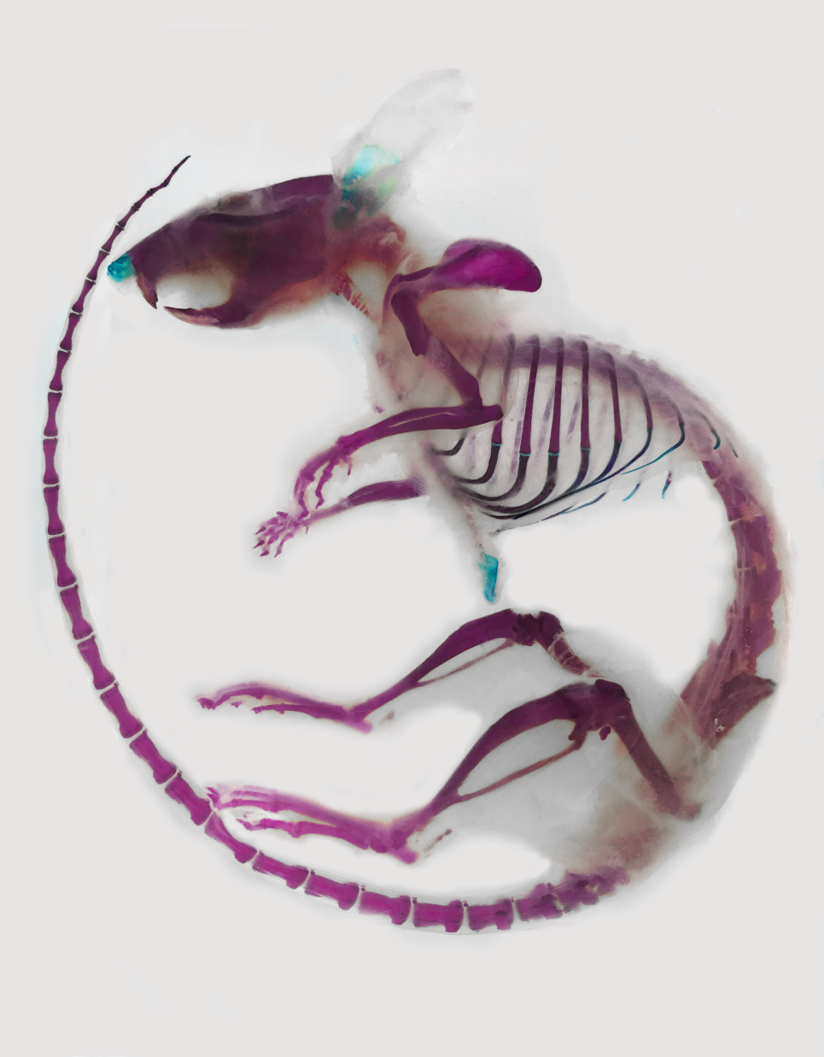
The purple color makes each skeleton part appear extra clear on the raw white background. From the tip of the tail, which is folded all the way around the body and up to the head, one can follow each vertebra in the tail up to the beginning of the spine, followed by the long bony hind legs. Gazing further, you reach the flimsy ribs, followed by the short forelegs, before one’s gaze eventually falls on the skull. The long front teeth reveal that the skeleton originated from a rodent.
The small skeleton, with its fascinating and enormous detail, is pictured in the photo, which has won second prize in the Danish National Research Foundation’s Photo Competition 2020, to which researchers from all fields of Danish research submitted their best research photos. The stripped skeleton, which appears in bright colors in the photo, is derived from an ordinary mouse of the species Mus musculus – also called a house mouse – which is commonly used as laboratory mice.
“The lab mouse in the photo has undergone a process called diaphonization, in which the animal is soaked in a fluid with the enzyme trypsin, which breaks down proteins. The enzyme has therefore consumed all the tissue, leaving only collagen, bone, and cartilage. The skeleton is then stained with a purple color that specifically binds to bone and a turquoise color that binds to cartilage. The collagen holds the animal together and can be seen as transparent markings. All in all, you are left with the detailed result seen in the photo after the end process,” explained the person behind the photo, Patricia S. Petersen, a staff scientist at the Novo Nordisk Foundation Center for Basic Metabolic Research at the University of Copenhagen.
Provides a detailed insight into the physiology of the animal
Diaphonization is a very common laboratory technique going back several decades. To this day, it is still widely used, as it is a relatively simple method for studying an animal internally and its physiology up close.
“In the past, people might have been sitting with a pile of bones trying to put them together to get an idea of an animal’s anatomy, but besides being laborious, it also does not provide an understanding of the musculoskeletal system. After diaphonization, on the other hand, you can actually move the animal’s bone parts around to observe how they normally function. So, you are able to create movements while studying them. Therefore, this method is worth gold for researchers like developmental biologists and zoologists,” explained Petersen.
Although Petersen and her colleagues in the research center at the University of Copenhagen also use the technique to study the physiology of laboratory mice, this is not what interests the research group the most. Diaphonization is really just part of the preparation work before experiments, as the researchers must be absolutely sure of the physical condition of the strains of mice used in the research. Mice strains are understood as mice that researchers follow for several generations or new types of genetically modified mice, where researchers, for example, have inserted entirely new genes or introduced point mutations in which a base in a gene is replaced by another base, to get the gene to code for something specific.
“We do a lot of metabolism studies, and in our research group, we focus on fat – especially brown fat – and then you can of course wonder why diaphonization is relevant to this. We use the method when producing new strains – that is, different genetic strains of mice that we use as research animals. It is a relatively simple method to check and make sure everything looks anatomically correct on our strains of mice,” explained Petersen.
Diaphonization is just one of the methods that researchers can use in the preparatory work of experimental mice. Other ways to closely study the mouse strains are cutting the mice open and examining them internally or observing their behavior and whether the animals are breeding as expected. In the end, the preparatory work is essentially done so that animals and time-consuming research do not go to waste.
“It is important that we know that what we genetically want to see in the mouse is as it should be before using it for research, because otherwise we risk a wrong starting point for the experiment, which can contaminate it and thus the results we get. It would be a disaster,” said Petersen.
Brown fat can be the key to less obesity
Petersen is part of Associate Professor Brice Emanuelli’s research group at the Center for Basic Metabolic Research. The research group focuses on research into type 2 diabetes and especially so-called brown fat. All humans have both white and brown fat, but unlike the well-known white fat that accumulates when we consume too many calories and store energy, brown fat is very different. It sits close to the nervous system, along the spine, at the neck, and around the kidneys, and instead of storing energy, brown fat burns energy to produce heat. Researchers around the world therefore hope that brown fat can be utilized to counteract overweight and lifestyle diseases related to overweight such as diabetes.
“In our research group, we look very much at thermogenesis – that is, the energy conversion in the body, and the idea that if brown fat is activated, then it can be used for something positive in relation to the metabolism. Can you, for example, counteract obesity using brown fat, or utilize the properties of brown fat to create a better treatment for diabetes? These are some of the questions we want answers to, and then we also look at the signaling pathway in the body, understood in the way brown fat so to speak ‘communicates’ to other parts of the body,” explained Petersen.
As a staff scientist in the laboratory, Petersen is part of the research staff, which you often don’t hear much about in science communication. This is despite the fact that these personnel play a large part in the world-class research that comes out of Danish universities every year. She has never had a great interest in writing scientific articles, but she loves the important legwork that is done in the laboratory, where her daily life offers a more practical approach. In her position at the research center, she is responsible for a number of smaller projects, but spends most of her time helping especially post-docs and other researchers on the research team with the hands-on part in the laboratory.
“I don’t have a great ambition to become a top researcher and write articles most of the time, because I really love the lab work. As long as I can work in the lab with something that interests me, which I find joy in, I don’t wish for more,” concluded Petersen.