Photo of aggressive brain cancer cells wins third prize in the DNRF Photo Competition 2019
-
The DNRF Photo Competition 2019: The three winning photographs in the Danish National Research Foundation’s Photo Competition 2019 have been found. In a series of three articles, the winners talk about their images and the research behind them. This article is about the third place winner, which this year went to Ph.D. student Johann Mar Gudbergsson. The articles about the first and second place winners can be found here and here. More information about the competition can be found here.
-
The panel’s review: The photo has a visual beauty that holds a sense of movement that underpins the invasive character of the depicted cancer cells. There is a demonic dimension in the motif that communicates the seriousness of the disease and, in that, the importance of the researcher’s work.
Panel:
- Christine Buhl Andersen, Director at Ny Carlsberg Glyptoteket
- Louise Wolthers, Research Manager/Curator at the Hasselblad Foundation
- Minik Rosing, Professor at the Natural History Museum, board member at the DNRF and Louisiana Museum of Modern Art
A photo of cancer cells in motion has been awarded third prize in the DNRF’s Photo Competition 2019. The cells originate from an aggressive type of brain cancer called glioblastoma; it is difficult to detect and defeat cancer cells that have spread to the tissue around the tumors. In his Ph.D. project, the winner of the third prize tries to map the cells’ movement patterns in the hope of contributing to the development of new types of treatment.
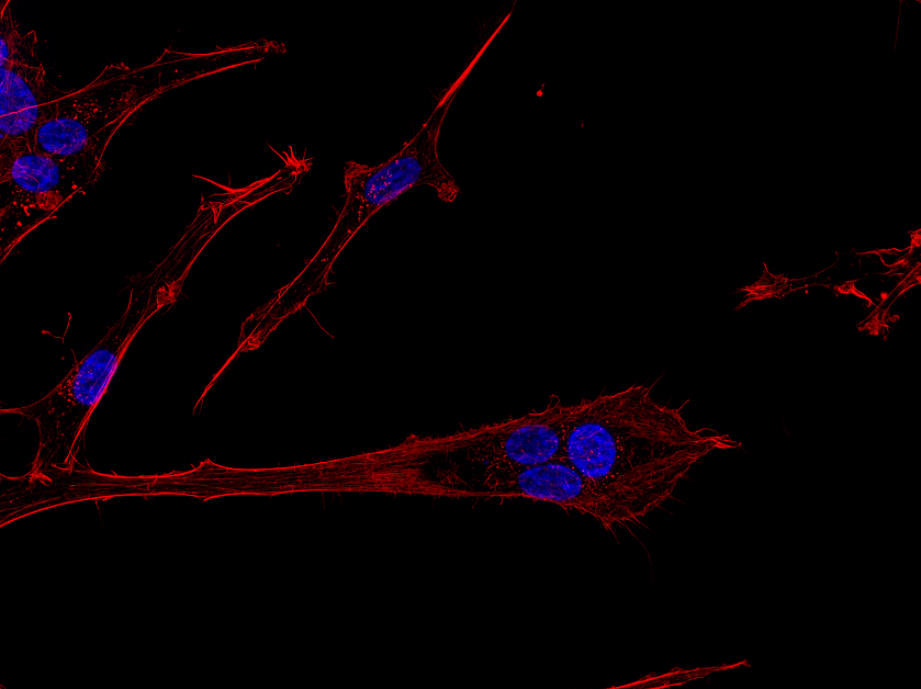
On a black background, thin, reddish fangs shoot out from blue spheres on the photo, which the selection panel has awarded third prize in the Danish National Research Foundation’s Photo Competition 2019. The image is a microscopic photo of aggressive brain cancer cells that are colored and processed to make the different cell components visible.
“The photo depicts brain cancer cells of the type glioblastoma, which is the most aggressive form of brain cancer we know of. The blue spheres are the cell nuclei, and the red is so-called actin filaments, a protein that forms part of the cells’ micro-skeleton known as the cytoskeleton. The cancer cells use actin filament to move, and as you can see in the picture, actin moves away from the blue-colored nuclei in an attempt to spread,” explained Johann Mar Gudbergsson, Ph.D. student at the Department of Health Science and Technology at Aalborg University and author of the picture.
When glioblastoma occurs in the brain, it typically develops into quite large tumors in several places. Since the brain is a very vulnerable organ that is central to our body and functioning, the position of the cancer also means that the disease is difficult both to treat and to examine.
“We do not really understand the mechanisms of glioblastoma yet, and part of the reason is precisely that the disease occurs in the brain, which is difficult to access. The location makes it problematic to perform a biopsy or examine the disease closely, as you can, for instance, with intestinal cancer or a tumor in the chest,” said Gudbergsson.
The only thing that can be done to treat this type of cancer is to operate, to remove as much of the tumors as possible.
Gudbergsson tries to model the invasive nature of the cancer
The problem with glioblastoma is that even though you remove the tumors, cells have often spread into surrounding tissue. As time goes by, new tumors will grow, which will require more surgery. These invading cells are the major problem for the treatment of this cancer type. For the same reason, there is no proven case where a person has been cured of glioblastoma.
One of the challenges is the so-called blood-brain barrier that separates the body’s circulating blood from the brain. The barrier consists of a dense layer of cells that protect the brain from harmful substances from the blood. But the barrier is so effective that neither targeted treatment drugs, for example, chemotherapy, can cross the barrier and attack the invading cells. At the location of the large tumors, the barrier is weakened, so that the drugs can attack the cancer. But in order to fight the invading cells in the tissue around the tumors, researchers have to search for other treatment methods. This is the reason that scientists like Gudbergsson would like to know more about the movements of the cancer cells and how the cancer spreads from the tumors.
“If we can learn to understand how the cancer cells move and thus model the invasive nature of the cells, then it is also easier to develop and test new forms of treatment that will hopefully work better than the treatments we have today,” said Gudbergsson.
(Video only available in Danish):
Models of cancer cells’ behavior can form the basis for new types of drugs
The picture, which has won the third prize in this year’s photo competition, is one of many thousands of microscopic images that Gudbergsson has photographed as part of the data set in his Ph.D. project, which he is scheduled to defend in a few months. In the project, the cells have undergone a treatment that enables Gudbergsson to insert and examine various proteins that play a role in the movements of the cells, including the aforementioned actin filament.
“This is, of course, basic research, but the intention is that the models I try to develop and characterize can be used when we try to treat the tumors in the laboratory or in animals. Here it is hoped that the models can help to create a fairly correct picture of the behavior of the cancer cells in humans, so that you can test new treatments as realistically as possible,” said Gudbergsson. He continued:
“Today, there are less than one percent of possible treatments that go through pre-clinical and clinical trials, but if we can learn to model the cancer’s mechanisms better from the beginning, then we may be more likely to develop new treatments and drugs that actually work in the end.”
Wants to make research his career
During his Ph.D. program at Aalborg University, Gudbergsson has collaborated with research groups from both the Technical University of Denmark and the University of Copenhagen, and as part of the project, he spent nine months in Copenhagen. After he defends his Ph.D. project in a few months, Gudbergsson will take a short break from research and concentrate on a couple of small companies of which he is co-owner. But there is no doubt in his mind that, in the long run, research is what attracts the young Ph.D. student.
“There is no doubt that I want to make a career in research and science; that is what I am passionate about. I have received a few offers, but for several reasons, this is not the time for me right now. I am in a transitional phase, and for the moment, I want to spend some time on my companies, which have been neglected lately. And at the same time, my girlfriend’s job opportunities also play into how the future will develop,” concluded Gudbergsson.
Read more about the first place winner here
Read more about the second place winner here
Further reading about this year’s competition can be found here